Research

Topics

Microtubule organisation and mechanosensation

Microtubule organisation and mechanosensation

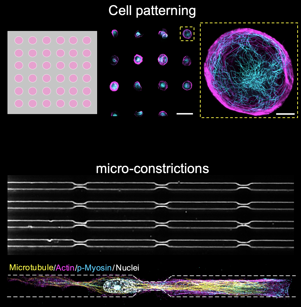
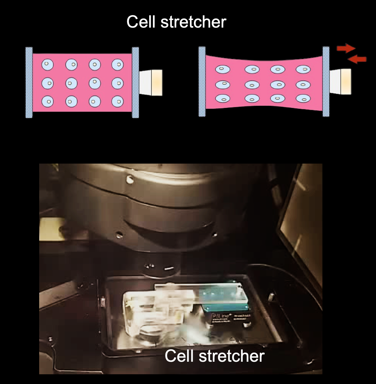
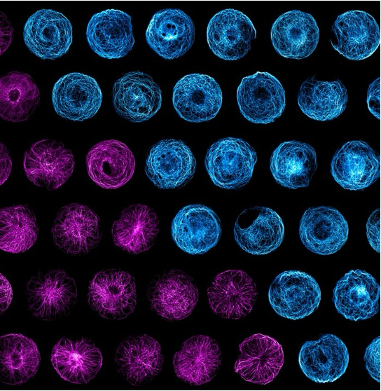
Microtubules (MTs) are stiff and dynamic polymers spanning the cytoplasm in mammalian cells. They are essential for maintaining vital living activities including mass transport, cell division, polarisation and ciliation. Our previous work (Nature Materials 2023) reported that the microtubule network can not only bear mechanical forces but rather respond to them, functioning as a microscale mechanosensor in living cells. MTs increase their stability to resist external forces by relocating plus-tip proteins along their entire length, and such a mechano-response is essential when cells migrate through constricted spaces. We are particularly interested in how MT mechanosensation coordinates with other cytoskeletons (e.g., actin and intermediate filaments) in cell migration or organoid morphogenesis models.

Preimplantation embryo — an in vivo model

Preimplantation embryo — an in vivo model

During mammalian embryonic development, a fertilized egg undergoes cleavages and develops into a blastocyst before implantation into the uterus. Embryos generate mechanical forces from the actomyosin machinery to guide blastomere compaction and internalisation. We are interested in how MT organisation and sensing function during this process and their potential links to key morphogenetic events, including compaction, apical-basal polarisation, asymmetric division, and cavitation. Addressing these fundamental questions on MT organisation will enhance our understanding of the basic rules of embryonic morphogenesis, which in turn, could help advance assisted reproductive technology and combat infertility.

Tools

We developed two categories of experimental tools to study cytoskeleton mechanobiology. The first category is for cellular mechanical perturbation, including cell patterning, cell stretchers, cell confinement/constriction. The second category is for cellular structure/mechanics characterization, including Traction Force Microscopy, and Expansion Microscopy (ExM). Particularly, we develop a protocol to visualise the ultrastructure of centrioles in mouse/human preimplantation embryos via ExM.

Cell patterningCell stretcherExpansion Microscopy