Lab Establishes U-ExM and Images Centriole Ultrastructure
research
We are excited to announce that CytoArchiLab has successfully established Ultrastructure Expansion Microscopy (U-ExM) in the lab.
Using this platform, PhD student Yang Wan imaged the centriole ultrastructure of human RPE1 cells, resolving the nine-fold symmetric microtubule architecture of the centriole on a conventional confocal microscope.
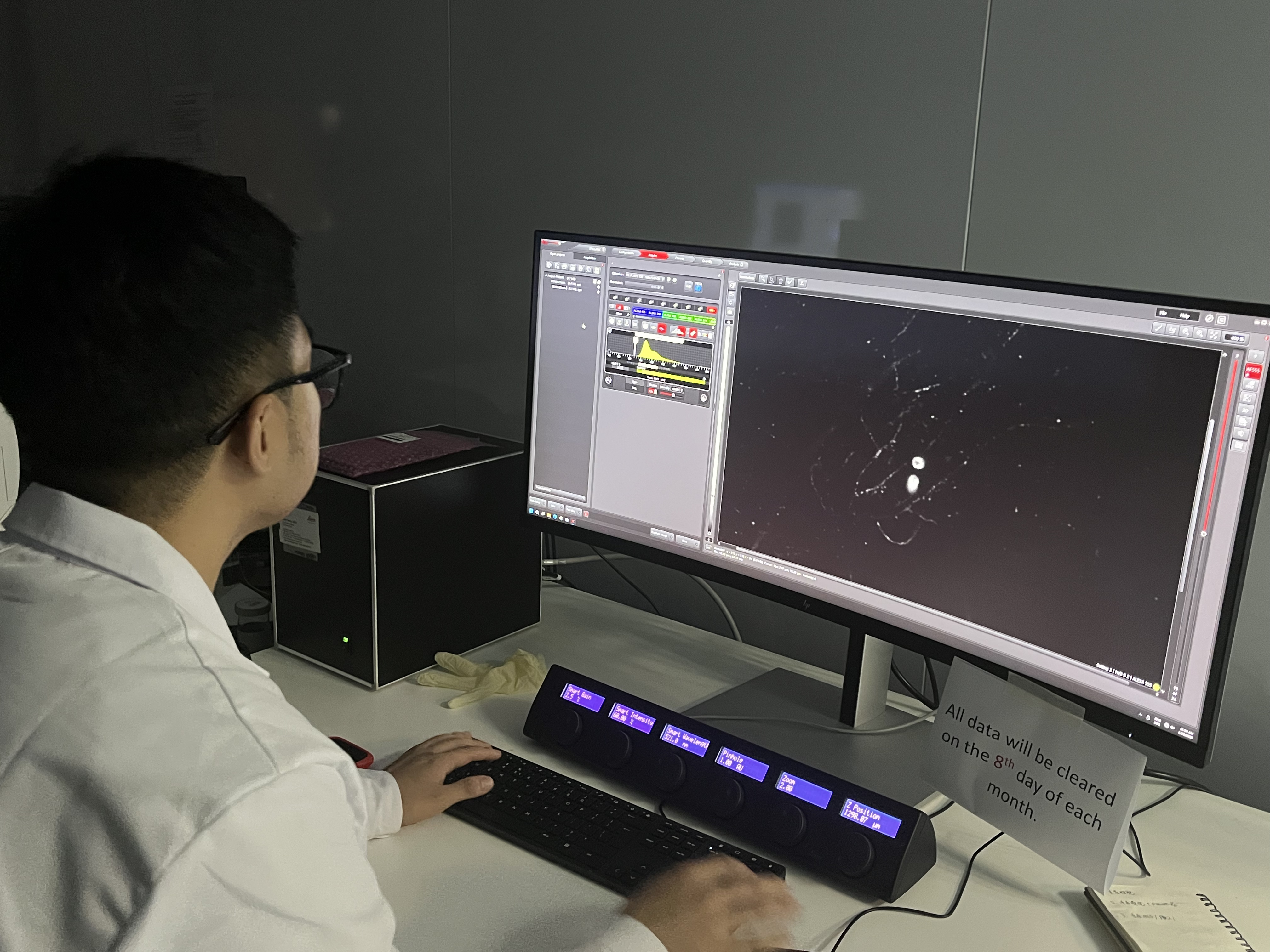
This establishes U-ExM as a core capability of the lab, paving the way for our studies on centriole assembly and cytoskeletal architecture across cell and embryo models.
Congrats to Yang Wan!